Best disease of the eye is also termed vitelliform macular dystrophy. As it is a type of macular dystrophy, this disease is caused by a fault in the gene. It can affect both males and females equally. Best disease is the inherited retinal disease in which macular affects the central part of the retina. It results in loss of central vision and the ability to perceive the colors.
The retina is the thin tissue lining at the back of the eye, which has rod and cone photoreceptors. They deal with the conversion of light into electrical signals. Then those signals transmit to the brain which interprets the signals into the vision. The best disease affects this visioning process because of macular degeneration which hinders the interpretation of electrical signals into vision.
Further, the best disease makes it difficult for the affected person to discriminate the details of the images seen by their vision.
How is the best disease of the eye inherited?
Best disease is inherited by one parent or both and it has happened when the mutation of BEST1 occurred. When the person who carries the gene with mutation of BEST1 paired with the other person having normal gene then there are 50 percent chances that the child is affected from the best disease.
It is because the child has one faulty gene from one parent while the other gene is normal and this is termed as an autosomal dominant inheritance. The BEST1 has happened in families who have a history of the autosomal dominant pattern of VMD2.
If the one parent has the faulty gene of the best disease but won’t transfer to the child then the child wouldn’t have the best disease. He wouldn’t be the carrier to pass on it to his offspring.
What is autosomal recessive bestrophinopathy?
There are rare chances in best disease to have a recessive autosomal inheritance which means, that the child has both the faulty genes from both the parents to be affected from best disease. This type of inheritance is known as autosomal recessive bestrophinopathy.
Genetic counseling helps a lot in family planning, genetic testing, and exploring the patterns of the gene mutation in the families to prevent it from passing to the next generations.
Symptoms of Best Disease of th eye
The progression of the best disease is varying person to person so the severity of the symptoms is also different in each affected individual.
Usually, the symptoms are found in early childhood or adolescence, whereas the growth rate of disease is generally slow which means the symptoms cannot be experienced throughout life or at a later age.
In adults, the symptoms of the best disease can be seen in between 30 to 50 years. If the progression in the disease is slow then the affected person may not experience permanent central vision loss or any significant loss of peripheral vision or discrimination of colors.
How does Best disease affect the eye?
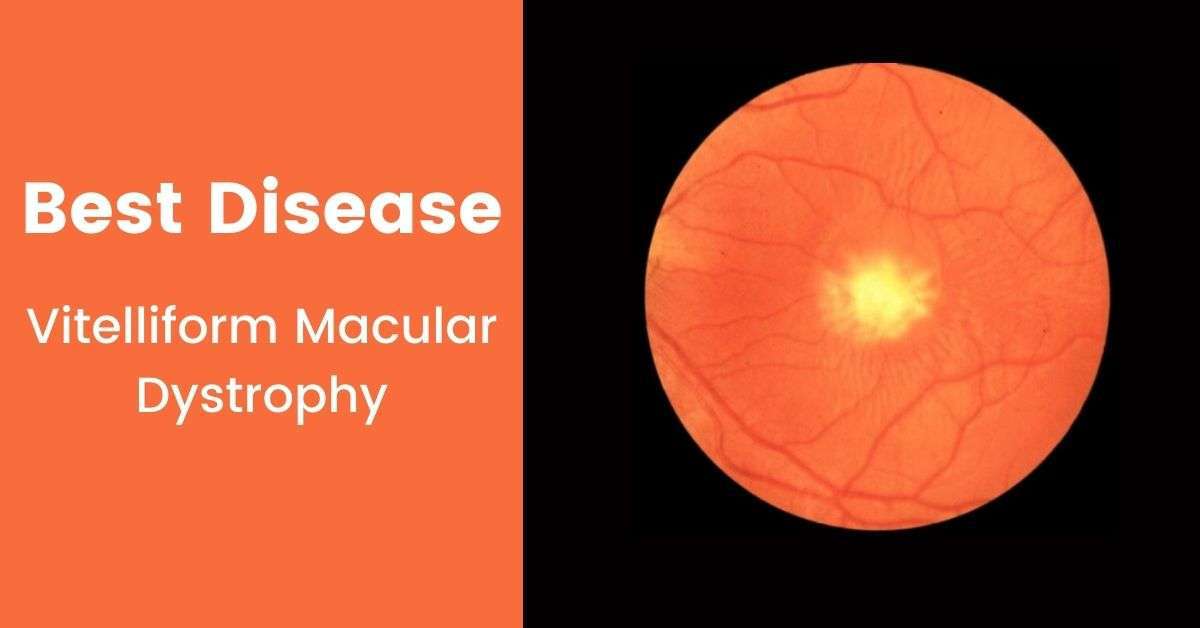
In the earlier stages, the symptom of the best disease is a bright yellow cyst, which is a sac filled with fluid, beneath the macula but under the retinal pigment epithelium (RPE). This retinal pigment epithelium is a thin layer of cells that provide support to photoreceptors. This cyst can be diagnosed by an eye doctor on examination as the cyst is seen as a sunny-side-up egg. This cyst can have no impact on visual acuity by having normal or near to normal vision and no effect is experienced on the peripheral vision.
Most of the individuals are having the cyst, generally experiencing the rupture of the cyst which is caused the yellow fluid to spread around the macula and the retinal pigment epithelium.
This fluid ends up with further vision loss. Retinas also have an accumulation of yellow flecks that is called lipofuscin which results in further vision loss.
Can you go blind with Best disease of the eye?
A significant number of best disease patients don’t experience any significant central vision loss throughout their lives. But still, doctors can detect it by conducting different diagnostic tests and checking the functionality of the retina and retinal pigment epithelium. Generally, the patient has a ratio of 20/100 deteriorating vision later in life.
Stages of Best Disease:
Many patients of the best disease don’t experience any early symptoms of it because even the child has the best disease but the child may never experience any effect in the sight until the optometrist somehow checks the macula.
But some cases experience issues with their sight but only their optometrist confirms the abnormality in the function of macula and confirmation of having the best disease.
Generally, the best disease happens from the age of 3 to 15 but a significant number experiences the issues in sight after 40 years of age due to the slow progression of this disease. The best disease has five stages for development in the retina and only the optometrist can confirm and tell about the stages because no stage causes the eye pain in the affected person; these stages are:
- Stage 1 – This stage of the best disease is the most initial one and does not affect the macula and generally the best disease is not directly affect the macula so there is no vision loss. In this stage, the layer under the macula is only affected otherwise the eye seems healthy.
- Stage 2 – The stage is known as the ‘vitelliform stage’. This stage is usually occurring between the ages of 3 to 15 years. In this stage, a blister has appeared in the macula but it has no effect or a slight effect on the vision. The optometrist can see that blister on the macula through the examination of the retina.
- Stage 3 – This stage is known as the ‘pseudohypopyon stage’. This stage can be experienced in teen aging and there is a slight change in the vision. The egg yolk-like blister on the macula can break through a further layer on the retina in the form of a cyst. The yellow-filled cyst can easily be seen in the retina by the optometrist.
- Stage 4 – This stage is known as the ‘vitelliruptive’. At this stage, some rupture can be seen in the cyst causing damage in the cells of the layer of the retina. Further, the person experiences a significant change in the vision like straight lines looks wavy whereas difficulty in reading small prints.
- Stage 5 – This stage is known as the ‘atrophic stage’. In this stage, the yellow material is getting out from the cyst and then disappearing but leaving the damaged cells in the layer of the retina. Now the patient experiencing serious issues with the reading as the words can’t be seen clear anymore.
What is choroidal neovascularization?
There are some cases of best disease only which is experiencing the development of one more stage during the atrophic stage and that stage is known as ‘chorodial neovascularisation (CNV).
In this stage, the eye tries to fix the damage by developing new blood vessels to recover the cells from the release of yellow material. But the new blood vessels lead to the development of scar tissue which further deteriorates the sight because the new vessels are leaky so they bleed.
Usually, the person with the best disease doesn’t experience a significant loss in sight till the development of stages 4 and 5. These stages are usually developed after 40 years. So patients don’t experience any issue with a vision till 40 years of age.
Diagnosis of Best Disease
The best disease can only be confirmed by eye health care. The optometrist is examined the retina and macula thoroughly to detect blisters or cysts present in the layer of the retina.
A proper eye examination can only tell about the severity of the best disease in the affected person and the growth rate of the disease that is damaging the macula. The optometrist further asked for the scans including:
- Optical Coherence Tomography (OCT)
- Electrooculography (EOG)
Optical Coherence Tomography (OCT)
The optical coherence tomography is the scan used to investigate the layers of the retina and usually, in the scan any blister or unusual particle can be seen if present in any layer of the retina.
Electrooculography (EOG)
Moreover, electrooculography is used to measure the response of the retina to the exposure of light as the retina deals with the photoreceptors that work on the sensitivity of the light on the retina.
The optometrist can also recommend genetic testing to confirm the diagnosis of the best disease and the requirement of further therapies and trials to stop or reduce the progression of the best disease. Genetic counseling also helps in understanding the gene mutation in the families which cause the best disease.
Is there any treatment for Best disease of the eye?
When it is about the treatment of the best disease then there is no proven treatment of it because the disease is caused by gene mutation and the reason for the gene mutation is still unknown. There is no proven treatment to stop this faulty gene mutation in the families of members having the best disease.
Chorodial neovascularisation and laser treatment
There is a very little number of patients of best disease suffering from the formation of the new blood vessels and this category is termed chorodial neovascularisation. These blood vessels are leaky and bleed further so laser treatment is used to remove these blood vessels to avoid further damage to the sight. The patient is also advised to avoid sports activities to reduce the chances of any hitting on the head because these blood vessels are developed due to head trauma.
Anti-VEGF drug injection
Moreover, anti-VEGF drug injection is used to stop or reduce the blood vessels to slow down the sight damage. The use of this anti-vascular endothelial growth factor medicine has only been prescribed by the optometrist because only the optometrist can see the position of the macula and the effect of blood vessels on the eyesight.
Further, gene therapy would be the potential treatment of the best disease according to the research. In this, a harmless virus is used to carry the normal gene in the retina and either stop the damage caused by the faulty gene in the cells of the retina or reverse the effect on the retina. Still, it is not a proven treatment but a ray of hope amid the fact that there is no proper treatment of this gene mutation disease.
How to cure Best disease of the eye naturally?
There is a long debate on it that there is no treatment of the best disease because it is caused by the gene mutation but there could be natural steps that are taken to reduce or stop the progression of the disease in the retina to avoid damage in the sight. Those natural cures could include the following:
- The patients of the best disease can intake key vitamins and minerals in the diet to stop or reduce the progression of the best disease to retain the sight as long as possible.
- Stay fit by having a healthy lifestyle through diet and exercise.
- Increasing use of carotenoids in the diet.
- Try to use eyewear to protect the eyes.
- Stop smoking.
Conclusion
The best disease is the rare hereditary eye disease which is caused due to the BEST1 mutation. The child has the best disease is usually carried the one faulty gene from the one parent and the one normal gene from the second parent.
The development of the best disease starts from the age of 3 to 15 but the significant effect in the central vision can be experienced after 40 years. The person experiencing the best disease generally has an issue with the central vision and the loss of color discrimination but the peripheral vision remains unaffected.
There is five-stage of the best disease and the sight remains unaffected till stage 4 and 5. The most prominent symptom of the best disease of yellow fluid-filled cyst in the macula which can easily be seen by the optometrist. The eye doctor can diagnose the best disease by examining the macula and retina through the optical coherence tomography scan and electrooculography scan.
Genetic testing is also useful in diagnosing the best disease. There is no proven treatment of the best disease is available.
Laser treatment and anti-VEGF medicine are used to stop or reduce the new blood vessels that are developed in very few patients with the best disease.
Gene therapy would be the potential treatment of the best disease according to the research and the ray of hope. As the reason for this gene mutation is yet to be disclosed.


