Human genes contain all the information regarding body development and working. One malfunctioning gene causes abnormal body development and results in a serious condition.
Such malfunctioning genes tend to pass down from generation to generation. And they disturb the order of our body. Such conditions caused due to gene error are called hereditary diseases or disorders. When we talk about the eye, we name such diseases as hereditary eye diseases.
Table of Contents
ToggleHereditary Eye diseases In Humans
There are certain eye diseases in humans that cause blindness. There are more than 350 eye diseases that have genetic causes. Not all hereditary eye diseases cause blindness. Many of these fall patients are in the low vision category, with impairment of visual functioning.
To make the topic better understand, we have classified these diseases according to infants, children, and adults.
Most common hereditary diseases that affect vision and may lead to blindness may include the next mentioned diseases.
Albinism, retinitis pigmentosa, retinopathy of prematurity, achromatopsia, corneal dystrophies, retinal dystrophies, Leber congenital amaurosis, glaucoma, keratoconus to name a few.
What Are Three Types Of Hereditary Eye Diseases?
For a better understanding, hereditary eye diseases can be classified as:
- Hereditary eye diseases that cause Childhood blindness
- Hereditary eye diseases that cause Adult blindness
- Rare Hereditary eye diseases that causes blindness
Hereditary Eye Diseases That Cause Childhood Blindness
In the eye, more than 60 percent of reported blindness in childhood is due to genetically caused diseases.
Low socio-economic conditions increase the rate of blindness due to hereditary disorders.
Autosomally inherited mode of disease transmittance, constitutes 22-52% of the genetic cause of blindness in children.
Mostly, the following hereditary and genetic causes, result in childhood blindness:
Retinitis pigmentosa affects a large number of people every year. It is one of those hereditary eye diseases of the retina where you go blind in both eyes. It may pass as an X-linked, autosomal dominant, or autosomal recessive trait.
Photoreceptors are the cells in the retina of the eye. These cells receive light and pass on signals towards the brain for visual perception. The faulty gene in retinitis pigmentosa causes the faulty formation of these photoreceptors which eventually affects vision.
Retinitis Pigmentosa develops gradually over the course of time and usually manifests after the age of 10. The first complaint patient presents with is he/she has difficulty in seeing at night. This is due to the degeneration of rods.
Rods are the photo-receptors responsible for night vision. The patients bump into things and have difficulty in finding objects. May also complain that he/she is not able to see at the periphery. A condition called tunnel vision.
This tunnel vision gradually converts into no vision at all. If your child is complaining about unusual vision problems, then take it seriously. Because it may be the symptoms of RP.
This disease has no cure at all currently. Only a diet better in vitamins can have some positive effects. Timely diagnosis and genetic counseling may help to reduce disease prevalence.
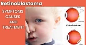
Cancer is among life-threatening diseases. Retinoblastoma is the cancer of the light-sensitive layer of the eye.
Children of age less than 5 years are more likely to develop this retinal cancer. The gene that causes Retinoblastoma is the RB1 gene. This gene is present on the long arm of gene 13.
When gene alteration affects both copies of the RB1 gene, it results in retinoblastoma. Uncontrolled retinal cell division causes tumor formation in one or both eyes.
If metastasis occurs, cancer may spread to the rest of the body and the child may even die.
As retinoblastoma is an inherited disorder, routine eye screening of parents and siblings of the child is also necessary.
Early detection is the key to preventing the child from both blindness and death. Red reflex seen during indirect ophthalmoscopy is the simplest technique to check the presence of retinoblastoma in the child’s eye.
The crystalline lens of the eye is a transparent structure. It is responsible for the bending of light and its focusing on the retina.
In congenital cataracts, the crystalline lens is cloudy by birth. Congenital cataract has a tendency to pass down from parents to their children.
So, if a parent or grandparent had a congenital cataract, new generations may have that gene too. How the fetus develops during pregnancy also has a relation with congenital cataracts.
Following are the risk factors that can result in the clouding of the natural crystalline lens during development.
- Maternal drug toxicity due to tetracycline antibiotics,
- Maternal infections during pregnancy like rubella, chickenpox, herpes simplex, etc.
Congenital cataract has various types, depending upon the area of lens clouding. If the opacity is central and blocks vision, then lens removal is necessary. When this cloudy lens stays in the eye, it will block the light rays. When the light rays do not reach the retina, it causes stimulus deprivation amblyopia.
The eye first becomes lazy and is not able to send proper signals to the brain. And gradually the patient may get blind.
Congenital glaucoma is among hereditary eye diseases causing childhood blindness. The ciliary process present in the posterior chamber of the eye produces a special fluid. This fluid is Aqueous humor.
The eye has a proper route for the drainage of aqueous humor. If anything obstructs the route, aqueous accumulates in the anterior chamber.
This increases the intraocular pressure (IOP), eye’s internal pressure. This condition is called glaucoma.
If by birth, any abnormal anatomical structure blocks this route of aqueous outflow, the pressure inside the eye increases immensely.
Due to raised IOP, pressure is exerted on all of the surrounding structures. These structures include the cornea, lens, vitreous.
It also damages the optic nerve head, especially the ganglionic fibers which carry electrical signals of vision towards the brain.
This damage first affects the visual field of the patient. And when the condition persists, irreversible loss of vision occurs.
Other names for Juvenile macular degeneration are Stargardt’s disease, Stargardt’s macular dystrophy, and fundus flavimaculatus.
There is a common question; does Stargardt’s disease leads to blindness? Well, yes it does. Mutations in the ABCA4 gene develop Stargardt’s disease and the disease travels as an autosomal recessive trait.
The gene in a healthy individual would clear away vitamin A by-products in photoreceptor cells of the macula. The macula is the central part of the retina responsible for crisp vision.
In macular degeneration, macular cells are depleted gradually. This happens due to the accumulation of vitamin A by-products (lipofuscin).
As a result, the eye progresses towards a loss of central vision, color vision, and stereopsis (depth perception). The damage once done is irreversible.
Due to its high genetic susceptibility, JMD is placed along with hereditary eye diseases.
Leber congenital amaurosis is a disorder of the retina caused by mutations in gene 14. This leads to severe visual impairment in infancy. Other associated conditions include high hyperopia.
In hyperopia or farsightedness; a person is not able to see near objects clearly. Nystagmus (jerky eye movements), extreme photophobia (increased light sensitivity), abnormal pupil response, and keratoconus (abnormally thin and cone-shaped cornea).
Usually, the disease travels in an autosomal recessive pattern like many other hereditary eye diseases. But when mutations occur in the IMPDH1 gene, the disorder is inherited as an autosomal dominant trait.
ROP is one of the most common causes of childhood blindness. The most important causative factors of ROP are premature birth of the child and excess oxygen exposure.
But, genetic susceptibility of ROP is also a factor that requires attention. On the basis of such researches, ROP is placed with hereditary eye diseases.
Premature infants are at a higher risk of ROP. Because when the gestational age is less than 7 months or birth weight is less than 1500 g babies have to be placed in incubators or ventilators.
Premature babies are placed in ventilators, where they are provided with oxygen. An oxygenated environment helps in their growth outside the mother’s womb. But prolonged oxygen provision has negative effects on the body of the child.
Due to excessive oxygen, extra new blood vessels grow on the retina of the eye.In medical terms, this condition is neovascularization.
These newly formed vessels may leak. That also causes scarring on the retina (light perceiving layer of the eye). This scarring detaches the retina from the underlying structure, resulting in blindness.
That is the reason, eye screening in premature infants is highly indicated. If the condition is screened out early, blindness can be prevented by anti-VEGF therapy and reducing the amount of oxygen.
Hereditary Eye Diseases That Cause Adult Blindness
There is a common concept that hereditary disorders only cause childhood blindness. No doubt, many of the genetic diseases manifest symptoms in early infancy or during childhood. But, there are some inherited and genetic disorders that manifest their symptoms in adulthood.
Children with such hereditary eye diseases are usually overlooked. That is because of the clinical features of such diseases.
Some of these disorders with genetic susceptibility and inheritance mode of transfer are:
Leber hereditary optic neuropathy (LHON) is that inherited disorder that causes vision loss in young adults. Why this disease affects males more than females, still remains an unanswered question.
Symptoms of LHON appear usually in the late teens or early twenties and rarely in childhood. That is why this disease has been placed under the adult blindness heading.
Central vision loss due to degeneration of optic nerve fibers is the most common feature.
Usually, this disease affects both eyes at the same time. But, if only one eye is affected, the other eye follows the track of the first eye within a week or two.
In some families, this disease manifests other symptoms as well. Like tremors (body shaking), cardiac conduction defects (abnormality of electrical signal conduction of the heart), movement abnormalities, and more.
LHON has a mitochondrial pattern of inheritance. It is the egg cell that contributes mitochondria to the developing embryo.
That is why this trait is also maternally inherited. Which means fathers cannot transfer the disorder.
The name of this hereditary eye disease is suggestive of the condition happening. ARMD is one of the leading causes of severe visual loss in people of age 60 years or more. ARMD is inherited from previous generations and gets worse over time.
There are certain predisposing factors of age-related macular degeneration. Like age, obesity, gender, smoking and heart diseases, etc. Macula the central portion of the retina of the eye is responsible for clear central vision.
In this condition, the macula starts degenerating. This causes first the clouding of the vision. Then gradually total loss of central vision, color vision, and stereopsis (depth perception of things) occur.
If symptoms of ARMD are recognized on time, disease progression can be helped.
Adult-Onset Primary Glaucoma
Adult-onset glaucoma shows the trait of family aggregation but strictly might not follow the Mendelian inheritance pattern.
This suggests that inherited risk factors have strong susceptibility to cause the disease. Other types of glaucoma like primary open-angle glaucoma, secondary glaucoma, angle-closure glaucoma do not have genetic relatability.
Glaucoma is a silent vision-killing disease. So, it is an urgent need that all the members of a family with positive glaucoma history undergo glaucoma screening.
Firstly peripheral vision is affected and gradually central vision also involves. The most common complaints are eye redness, pain, and photophobia.
Gonioscopy and tonometry are two reliable methods for the early detection of glaucoma.
Gonioscopy is a technique for checking contour and the shape of the angle of the anterior chamber.
Tonometry is a technique for checking the eye’s internal pressure due to the fluid in the eye.
Rare Hereditary Eye Diseases That Cause Blindness
Rare eye diseases affect less number of people but have irreversible effects on vision. These conditions are usually syndromes.
Syndromes word is used for a group of disorders happening together and each part of the body manifests unique symptoms.
In the eye, many genetic and inherited syndromes result in blindness. Some of these rare eye conditions are:
Usher syndrome mainly affects hearing due to the malfunctioning of the inner ear and auditory nerves.
Due to the involvement of the inner ear, the patient experiences balance problems too.
With regards to the eyes, retinitis pigmentosa occurs in the patient with Usher syndrome.
There are three types of Usher syndrome:
- In type 1, the patient experiences profound hearing loss and extreme balance problems. Vision problems begin in early childhood.
- In type 2 of Usher syndrome, hearing inability varies from moderate to severe. Vision problems usually do not start till the late teen years.
- In type 3, until puberty, the patient experiences no problems in hearing and body balance. Patient is just like any other ordinary individual. But vision problems are progressive since birth.
Usher syndrome has no cure, but gene therapies might have a positive effect.
Best disease is a type of macular dystrophy namely vitelliform macular dystrophy. The best disease affects 6 in 100000 people and passes down from generation to generation in an autosomal dominant pattern.
It is grouped with hereditary eye diseases because it causes a material build-up in the macula since birth.
During the time period when the child grows into an adult, the build-up gradually enlarges. That way the macula has already been damaged beyond treatment. No remedy can reverse vision loss.
The best disease affects both eyes but the extent of vision deterioration might not be the same in both eyes.
The disease shows symptoms later in life and sight loss may occur even after 50 years of life.
Mutation in the gene named BEST1 causes Best disease. The best disease has no cure to date.
Bietti’s Crystalline Dystrophy
Bietti’s crystalline dystrophy is one of the rare autosomal recessive hereditary eye diseases that causes blindness. People with Asian lineage are at the most risk of having Bietti’s crystalline dystrophy.
The disease starts developing after birth but symptoms manifest in early adulthood.
Yellowish-white crystalline lipid deposits accumulate in the cornea (sometimes) and the retina (most of the time) of the affected person.
The patient’s first complaint is cloudy central and peripheral vision or night blindness.
Choroideremia
Choroideremia affects all the retinal layers of the eyes. This is one of the hereditary eye diseases, that starts showing symptoms in childhood due to the damage to the retina, retinal pigment epithelium, and choroid.
The mutations in the gene CHM cause choroideremia. There is a gene in the body called the CHM gene. CHM gene is responsible for the production of a special protein. This special protein indirectly controls intracellular trafficking.
Intracellular trafficking is basically the movement of organelles within a cell. When this escort protein is absent, premature cell death occurs.
This whole process when occurs in the retinal cells, progressive vision loss has resulted. Choroideremia passes down the generations in an X-linked recessive pattern.
That is the reason, that this disease affects males more frequently than females. Other names for choroideremia are choroidal sclerosis and progressive tapetochoroidal dystrophy.
Conclusion:
Hereditary eye diseases, either manifested in childhood or in adulthood, progress gradually.
Usually, hereditary eye diseases cause blindness because the patient and his family have no awareness about the condition.
It is important to diagnose these diseases at an early stage. This leads to adequate management and treatment.
Families that have hereditary eye diseases running along with generations, should focus on routine eye examination of every member.
After a child’s birth, parents should always pay close attention to their child’s response and behaviors.
If the parents notice anything unusual, they should take their child to the nearest eye facility immediately.
In the cases of premature births, routine eye check-ups are mandatory. ROP, retinopathy of prematurity, can be detected early via fundus examination. This can prevent permanent vision loss.
Genetic counseling is one other major factor that helps. People have no idea that they may be the carrier of the faulty gene.
In cousin marriages, mutated gene shuffles but always remains there to cause malfunction.
So, it is better to avoid cousin marriages when the family is already suffering from a hereditary disease.
In adults, slit-lamp bio-microscopy, ophthalmoscopy, tonometry, and visual field examination are necessary for every follow-up session. So that, the doctor or primary eye care physician can effectively manage the condition.

The latest research and updates straight to your inbox !
Join our subscribers for exclusive access to our monthly newsletter related to optometry