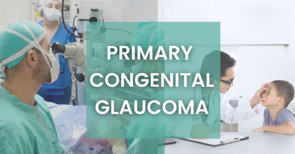
Are you looking for guidance related to primary congenital glaucoma?
In this article, we have discussed major aspects of this disease. This article will be very helpful for the parents of infants.
As early diagnosis plays a vital role to cure this disease effectively.
Table of Contents
ToggleWhat is Glaucoma?
Glaucoma is an eye disease in which high pressure damages the optic nerve. This nerve acts as a linkage between the eye and the brain.
Our eyes produce a fluid called aqueous humor. If this fluid does not drain out properly, it will increase the pressure in our eyes. This pressure is called interocular pressure(IOP).
This IOP directly affects the optic nerve. The optic nerve consists of small tiny nerve fibers.
If IOP damage this fiber, it will cause a blind spot in the vision. If all fibers are damaged completely, our vision will be a complete loss.
What is primary congenital glaucoma?
Glaucoma is a normal eye disease in middle age.
In some cases, glaucoma happens in the childhood stage. This is a very uncommon state in childhood.
It causes severe damage to the vision of an infant. This type of glaucoma is known as congenital glaucoma.
In congenital glaucoma, there is a genetic birth defect in the angle of the eye. This fault hinders the flow of aqueous humor.
Consequently, interocular pressure rises and it harms the optic nerve directly.
If congenital glaucoma occurs without any specific reason, we can call this condition primary congenital glaucoma. This is one of the hereditary eye disease causing childhood blindness.
Primary Congenital Glaucoma Inheritance
In most cases, PGC occurs in infants with no family history of the disease. 10% to 40 % of cases are with a known family history of PGC.
The researchers found that the pattern of inheritance is autosomal recessive (Autosomal recessive traits pass from both parents onto their child).
But in some cases, researchers have also observed the autosomal dominant inheritance pattern (Autosomal dominant traits pass from one parent onto their child).
Types of primary congenital glaucoma
PCG has the following types, on the basis of onset:
1)True congenital glaucoma/ newborn onset (0-1 month)
In this type of glaucoma, the researcher observed optic ocular expansion already present in the first month.
They believed that interocular pressure elevates before birth by itself. These cases are about 25 percent.
2)Infantile glaucoma/ infantile onset (>1-36 months)
This type is present between 1 to 36 months and is about 65 percent of all cases.
3)Juvenile glaucoma/late-onset or late-recognized (>36 months)
This glaucoma is present in the age group after three years but before the adult stage. It is about 10 percent of the total cases.
Symptoms of primary congenital glaucoma
PGC has the following symptoms:
- Discomfort in bright light ( Photophobia)
- abnormal contraction of the eyelid muscles (Blepharospasm)
- Excessive tearing in the eyes.
- Redness in the eyes
- Swelling of the eyelids
- Presence of Cloudiness of the cornea
- Abnormal or enlarged eyeballs
Causes of primary congenital glaucoma
PGC has the following main causes:
- Elevation in interocular pressure
- Enlargement of the globe (Bupthalmos)
- High aqueous humor fluid in the eye
- Opacification or whitening of the cornea
- Defects in the ocular angle of the eye by birth
- Issues in the development of cells or tissues
Diagnosis of primary congenital glaucoma
In the case of congenital glaucoma, we cannot ignore the role of consanguineous marriage or cousin marriage.
As discussed earlier the inheritance pattern of PGC is autosomal recessive but in some cases, it may be autosomal dominant.
To diagnose the presence of PGC, the ophthalmologist first examines the eyes of the patient.
Detailed Clinical Examination of Eye
In this detailed examination, the ophthalmologists make the following checkup:
- Ability to follow or fixate the light
- Detailed inspection of Sclera (White surface of the eye)
- Thorough check of Cornea (the transparent part of the eye that covers the front portion of the eye)
- Anterior chamber check-up of the eye
- Iris inspection (the colored part of your eye)
- Pupil examination (the black circle in the center of the iris)
- Lens Quality
- Optic disc checkup
- Intraocular pressure Checkup
Evaluation of Clinical Examination
If an ophthalmologist found anything suspicious during the examination of the eye for PGC, they carried out further inspection of that part under anesthesia:
- Corneal Examination/Measurement
In the corneal examination, the ophthalmologist measures the diameter and evaluates the anterior section of the cornea by using calipers and slit lamps.
- Ophthalmoscopy
In ophthalmoscopy, the ophthalmologist evaluates the dilated fundus in detail. He will make a complete check-up of the optic disc. He will also check if there are any abnormal vessels causing the problem.
- Intraocular pressure (IOP) Check-Up
The ophthalmologist will check the IOP by using a Schiotz or handheld Perkin’s applanation tonometer, or a pneumotonometer/tonopen. IOP will range from 30 to 40 mmHg, due to anesthesia.
- Gonioscopy
In gonioscopy, the ophthalmologist will check the drainage angle of the eye by using Koeppe’s lens. If the angle is normal, it will help out aqueous humor to drain out properly.
- Pachymetry
In pachymetry, the ophthalmologist will measure the thickness of the cornea.
- Axial length
The ophthalmologist will measure the axial length of a child by using ultrasound. Axial length ranges from 18 mm at birth to 22 mm till 2 years.
- Cycloplegic retinoscopy
In cycloplegic retinoscopy, the ophthalmologist will check the refractive errors in the patient.
Treatment of Primary Congenital Glaucoma
The treatment of primary congenital glaucoma depends on the age and the severity of the condition of the patient at the time of diagnosis.
Although surgery is the basis of treatment, a medicinal treatment may be vital in a few patients depending on their condition.
Angle surgery, either goniotomy or trabeculotomy, is the gold standard therapy for primary congenital glaucoma because it reduces IOP by increasing aqueous outflow.
If angle surgery fails, trabeculectomy with mitomycin C or a glaucoma drainage device should be tried.
In stubborn situations, cycloablation with diode cyclophotocaogulation, Nd YAG, or cryoablation can be undertaken.
Topical or oral antiglaucoma medications are used to reduce corneal edoema, manage IOP, and improve visualization for goniotomy.
Medical Treatment
Ophthalmologists use this method for the following reasons:
- To lower the IOP of the patient
- To decrease corneal edema (swelling of the cornea)
- To enhance angel visualization of the eye
- Waiting for surgery
- To get the patient fit for anesthesia
Ophthalmologists refer to topical medicines and drugs in this medical treatment. This medical treatment does the following:
- Can reduce aqueous humor production.
- Can increase aqueous humor discharge.
1) In case of reduction in production in aqueous humor, ophthalmologists suggest Alpha-adrenergic agonists such as brimonidine and apraclonidine, beta-blockers such as timolol, betaxolol, and levobetaxolol, and carbonic anhydrase inhibitors such as brinzolamide are examples of medications that reduce aqueous humor production.
2) In case of an increase in aqueous discharge, contain parasympathomimetic drugs or prostaglandin analogs like travoprost.
Some above-mentioned drugs may cause respiratory depression or sleepiness in children.
Surgical Treatment
Ophthalmologists divide surgical treatment into:
- Angle Procedures
- Filtration Procedures
First we’ll discuss angle procedure:
1)Angle Procedure
Angle procedure rises the aqueous humor discharge lowering the pressure. The angle process is further divided into
- Internal approach using goniotomy
- External approach – Trabeculotomy
Internal approach using goniotomy
In the internal approach, the ophthalmologists used gonio lens to carry out the procedure.
This method requires a somewhat clean cornea that allows for clear observation of angle structures. Success rate is between 70% to 90%.
External approach – Trabeculotomy
This is effective in circumstances when the cornea is blurry owing to clouding or when Goniotomy treatments have failed. This technique is performed on the 12 o’clock incision, opening the trabeculum 120 to 180 degrees.
A novel modification approach with a prolene suture has been reported. This enables 360-degree trabeculotomy and permits the full angle to be opened in a single sitting. According to the research, trabeculotomy reduces intraocular pressure by 75 to 90%.
2)Filtration Procedures
Filtration procedures include following:
Trabeculectomy
Trabeculectomy procedures alone are not commonly performed in PCG due to low success rates due to children’s exaggerated healing responses.
In PCG patients, success rates ranging from 52 to 82% have been observed following anti-metabolite-assisted trabeculectomy surgeries.
Deep sclerectomy
This method entails raising a partial-thickness scleral flap and removing the exterior component of Schlemm’s canal and the outside part of the trabecular meshwork, including juxtacanalicular tissue, without fully piercing the eye.
Glaucoma Drainage Devices (GDD)
The role of GDD as the main procedure in primary congenital glaucoma is restricted.
In circumstances when primary angle treatments have failed or in advanced refractory cases, GDD implantation is recommended in primary congenital glaucoma.
One year after surgery, studies demonstrate a drop in intraocular pressure of 28 to 49%.
Combined Trabeculotomy and Trabeculectomy (CTT)
When the preceding trabeculectomy fails or there is trouble cannulating the Schlemm canal, this surgery is attempted.
The trabeculectomy is combined with the trabeculotomy in this procedure. Surgical peripheral iridectomy is used to remove a block of tissue from the sclera. When taking mitomycin C, use caution.
Cyclodestructive Procedures
This technique is only for eyes with a poor or non-existent visual prognosis. Only around 30% of these situations are successful.
A better and safer option is a modern modification in the form of a micropulse transscleral diode laser.
Conclusion
The overall prognosis of primary congenital glaucoma is determined by the time and severity of presentation, the age of onset, and the clarity of the cornea.
In many circumstances, early identification and care are critical to maximizing visual potential.
Corneal edoema and optic nerve head abnormalities are reversible if treated promptly.
Children must be checked for and treated for any related refractive errors caused by axial elongation of the eyeball.
Intraocular pressures and the optic nerve head must be checked on a regular basis for the rest of one’s life. Late presentations may result in vision-threatening consequences.
Research from the United States found that 90.3% of patients did not advance after sufficient therapy after one year, 83.1% after five years, 70.8% after ten years, and 58.3% after 34 years.
As a result, the need for effective patient treatment and follow-up is highlighted.
Another research found that angle surgeries were 90% effective in patients between the ages of 2 months and 1 year, compared to 50% in infantile, late-onset, or late-recognized instances.

The latest research and updates straight to your inbox !
Join our subscribers for exclusive access to our monthly newsletter related to optometry